
Pregnancy Anatomy
When a woman is pregnant and her body changes every day. The physical changes begin occurring almost immediately with release of hormones. These hormones are likely responsible for mild to extreme fatigue and nausea as well as breast tenderness and swelling.
Hormones
HCG (Human Chorionic Gonadotropin) is produced by the embryo and later the placenta and helps maintain the ovary’s ability to release progesterone. Estrogen promotes the growth of the uterus, stimulates duct system and blood supply in the breasts and influences water retention, skin pigmentation and subcutaneous fat buildup.
Progesterone has a number of roles in the pregnant body. It relaxes the uterus and inhibits contractions therefore its levels change throughout pregnancy. It relaxes the walls of blood vessels to help maintain low blood pressure and relaxes the walls of the bowels and stomach to help with nutrient absorption. Progesterone also stimulates the production of the hormone Relaxin.
Pregnancy Anatomy Photo Gallery
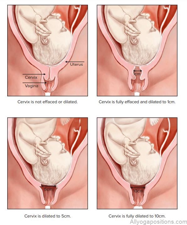
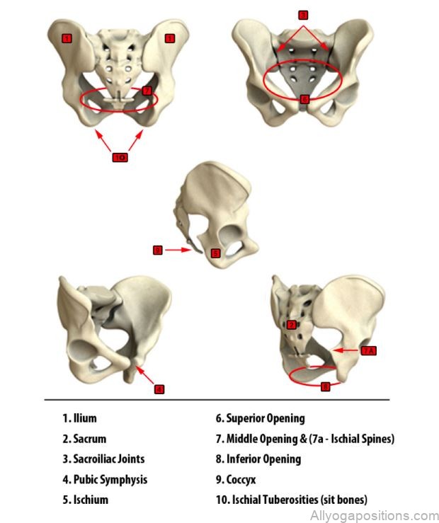

Relaxin lubricates the joints and softens connective tissue allowing the pelvis to open up to accommodate the baby. However hormone is not specific to the pelvis and it affects every other joint as well and can create instability in the joints this is why you hear to many pregnant women complaining of carpel tunnel syndrome. Therefore, you need to make sure your students take care not to overstretch. The Uterus The Uterus starts out about the size of a fist and is tilted slightly forward in most women above the bladder and in front of the rectum and bowel. The entrance of the uterus is called the cervix.

The uterus is made up of 3 layers: Inner layer called the endometrium is a mucus lining which makes up the very inner wall where the placenta attaches. This layer is what is shed during menstruation. Middle layer called the myometrium, a combination of crisscross and lengthwise muscles that grow to 10x in length and 3x in width during pregnancy. The fundus is the group of muscles included in the myometrium that runs from the cervix to the top of the uterus and is measured to help determine gestational age. These muscles contract and shorten during labor making the cervix shorter and helping the baby move down. Outer layer is called the perimetrium, it is loose tissue that surrounds the uterus and separates it from the intestines.
The Pelvic Floor
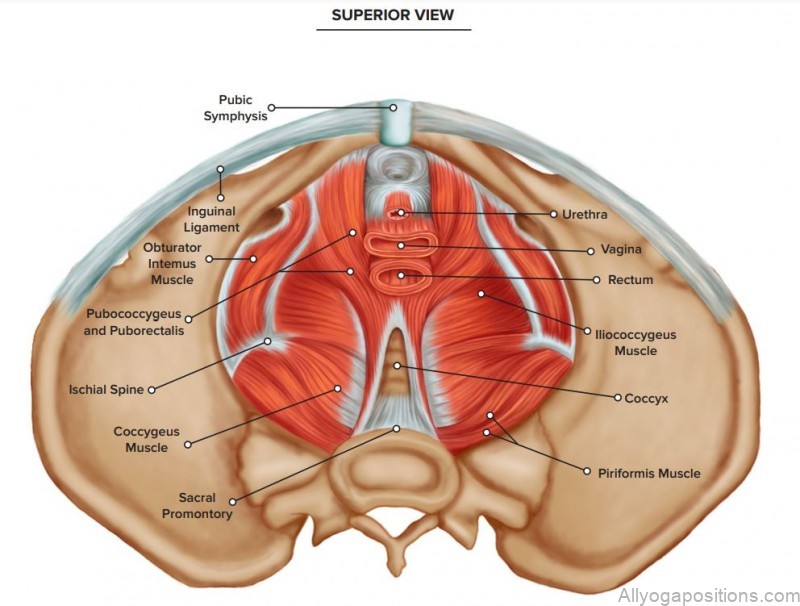
The pelvic floor muscles are a hammock of muscles connected to the pubic bone and backbone that hold up the uterus, bladder and intestines. These muscles help control urination and bowel movements and help guide the baby through the birth canal during the pushing phase of childbirth. Because of the amount of weight the pelvic floor has to accommodate during pregnancy and the stretching these muscles do, they can easily become weak, thus causing urinary or anal incontinence. Pelvic floor exercises are a pivotal part of prenatal health.

Table of Contents
Maybe You Like Them Too
- Tranquil Touchpoints Yoga for Inner ConnectionA gentle yoga practice to help you find peace and balance in your mind, body, and spirit.
- Yoga for Rehabilitation A Holistic Approach to Healing the Body
- Tolasana Yoga Pose A Guide to This Inverted Backbend
- Yoga for PTSD Recovery A Mindful Way to Heal
- Tittibhasana Yoga Pose A Guide to This Inverted Arm Balance